



An MRI scan for back pain is needed when it is associated with leg weakness, numbness, bladder or bowel symptoms, fever, weight loss, recent injury or when it persists for more than six weeks despite treatment. Most back pains of minor nature heal without scans.
Your back has been hurting for quite some time, and now you are hearing about an MRI! Do you really need one?
Back pain affected 619 million people globally as of 2020 and is predicted to rise to 843 million by 2050. Nevertheless, the majority of these patients do not require a scan at the outset. Here are some of the signs that can help you understand when an MRI for back pain becomes necessary. You will also learn about the differences between an MRI and an X-ray and the next steps to take if you experience any of these symptoms.
Does Every Back Pain Require an MRI Scan?
No, not every back pain requires an MRI scan. Most back pain occurs because of muscular or joint problems. According to the World Health Organization, there is about 90% prevalence of non-specific low back pain, which means there are no structural issues seen on scans of such patients.
Why is most back pain non-specific?
The muscles, joints, and ligaments in the lower back region are subjected to high levels of stress throughout the day due to sitting and physical work. Usually, this type of pain goes away on its own after light exercises. Physiotherapy and over-the-counter medications can assist in the healing process. However, an MRI scan is unnecessary in this situation.
What are the reasons scanning too early can be misleading?
A large number of healthy people also show some disc bulging or signs of degenerative change on MRI scans. As a result, getting an MRI scan performed before consulting a medical professional can cause you to concentrate on the wrong issue. A physician will correlate the findings from the MRI scan with your symptoms and discover the exact cause of your discomfort.
Signs That an MRI for Back Pain Is Necessary
A physician recommends an MRI scan when the symptoms suggest a problem with the spinal cord, intervertebral discs, or spinal nerves. For example, if you have any of the following symptoms, you should consult one of the best spine hospitals in Chennai:
- Shooting pain from the buttocks down the leg, accompanied by tingling and weakness, is an indication of sciatica, slipped disc, or disc prolapse.
- Pain that occurs at night or does not respond to rest.
- Pain that persists for more than six weeks despite therapy and medication.
- Pain that commenced after an accident or injury.
How soon you need to get an MRI depends on the severity of your symptoms:
| Symptoms | Possible Cause | Next Steps |
| Numbness in the groin or buttocks, as well as loss of bladder or bowel control | Cauda equina syndrome | Seek emergency medical attention |
| Back pain with fever | Spinal infection | Get evaluated the same day |
| Back pain with unintentional weight loss or history of malignancy | Tumour or metastasis | Schedule an MRI scan immediately |
| Minor leg numbness, which is resolving | Nerve irritation | Consult your doctor and reschedule the MRI scan |
In addition to the symptoms listed above, persons over the age of 50 are more likely to have long-term back discomfort, according to the World Health Organization. As a result, they should get an MRI scan earlier.
When Is an MRI Necessary for Lower Back Pain?
Pain that persists for more than six weeks
If your lower back pain is not resolving after approximately six weeks of treatment, an MRI scan can help identify the underlying cause. An MRI in such situations allows you to rule out the possibility of disc prolapse, sciatica, or any other significant problems.
Sciatica and leg pain
Pain that radiates from the buttock down the leg indicates sciatica. In addition to the typical sciatic nerve pain, there may also be numbness or weakness in the legs. An MRI scan helps in localizing the affected spinal nerve thus isolating the causative disc protrusion.
Before injections or spine surgery
An MRI scan is advised before injections or spine surgery. It helps the surgeon in localizing the precise location to be treated.
What Does an MRI Scan Reveal for Back Pain?
An MRI scan uses strong magnets and radio waves to capture images. Unlike X-ray machines, MRI scans do not expose you to harmful radiation. With an MRI, it becomes possible to view delicate tissues that an X-ray cannot penetrate. An MRI scan can reveal the following:
Slipped disc and disc prolapse
The MRI scan will show whether any disc has ruptured, protruded, or bulged. Your report may indicate bulge, protrusion, or herniation with varying degrees of severity. These terms imply different levels of disc damage. The amount of pressure being exerted by the bulging disc on the spinal cord and nerves can also be determined in some instances.
Does an MRI scan show nerve compression?
Yes, an MRI scan can show whether a spinal disc, ligament, or bone spur is putting pressure on the spinal cord or a nerve root. MRI scans are often recommended for patients presenting with leg numbness and weakness to identify the causative nerve compression.
Spinal canal narrowing, infection, and tumours
An MRI scan can also be used to diagnose spinal stenosis, infection, or tumours. Such conditions require immediate attention and therefore, the sooner they are detected, the better.
MRI vs X-ray for Back Pain: What is the Difference?
X-ray and MRI scans are both essential diagnostic techniques. The difference between an MRI and an X-ray lies in the information each provides:
| X-ray | MRI |
| Helpful in diagnosing fractures and the status of the bones | Helps visualize disc prolapse, nerve compression, and spinal cord damage |
| Exposes you to low levels of radiation | Does not expose you to any radiation |
| Quick scan: takes a few minutes | Longer scan: takes about 20 to 40 minutes |
An X-ray is generally the first test to be taken after an accident because it is a quick procedure that provides useful information about the state of your bones. An MRI scan, on the other hand, is more appropriate when there is a suspicion of nerve or disc injury.
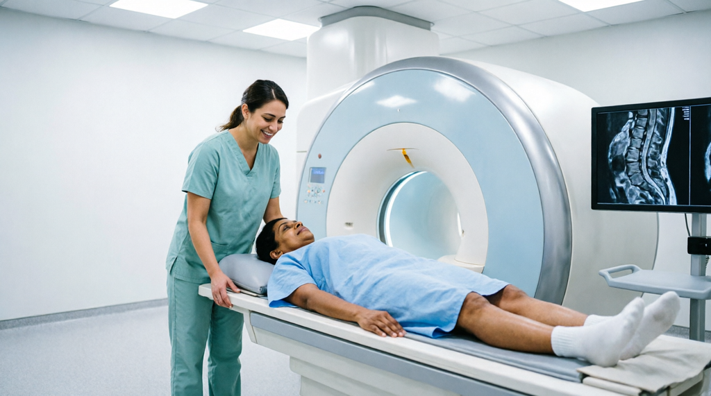
What to Expect During a Back MRI
Preparation before an MRI scan
Before the commencement of the procedure, inform the technologist about any metallic implants or devices in your body, such as pacemakers, surgical clips, or cochlear implants. Metallic objects must be removed before the scan commences.
MRI scan procedure
You will lie on a table that slides into the machine. Earplugs or music may be provided to help you relax during the procedure. The MRI machine makes some rather uncomfortable noises, thus it is critical to remain still during the procedure. A back MRI scan procedure takes about 20 to 40 minutes.
If you feel claustrophobic, speak with your doctor about the options he or she has to make you feel more comfortable during the procedure.
What happens after a back MRI scan?
You can typically return to your regular activities right after the scan. The radiologist will go through your results and provide you with a copy of the MRI report for your doctor to review.
How to Read an MRI Report for Back Pain
Slipped disc terminology
Doctors classify a slipped disc using the following terms:
- Bulge: The disc stretches out slightly beyond the edges of the vertebrae.
- Protrusion: The disc pushes out farther and begins to bulge.
- Herniation: The disc material goes beyond the protrusion and begins to bulge.
Stenosis of the spinal canal
Spinal stenosis is a condition in which the space for the spinal cord is narrowed due to swelling of the ligaments or reduction in spacing between vertebrae. Your report may state that spinal stenosis has been discovered.
Does a “bulging disc” always mean that it is the cause of pain?
No, it does not. There is typically nothing to worry about if the MRI scan shows only a slight bulge. A bulging disc is most likely to be related to the discomfort if it is moderate or severe. Remember that the MRI scan findings should always be interpreted in conjunction with the symptoms.
Treatment Following an MRI Scan for Back Pain
Physiotherapy and pain management
The majority of patients with back pain are treated with physiotherapy, medications, and pain management. If conservative therapy fails to relieve your symptoms, you can always explore other treatment options such as injections with our pain management experts.
When is surgery required for back pain?
Most of the time, surgery is not recommended as the first-line therapy for back pain. Surgeries are typically reserved for patients with progressive neurological symptoms, bowel or bladder dysfunction, or significant pain that is not controlled with conservative therapy.
Choosing a Spine Specialist in Chennai
When choosing a back pain specialist in Chennai, you should look for the following:
- A specialist who examines before recommending an MRI scan.
- A spine expert with orthopaedic expertise.
- One who has an in-house diagnostic centre, eliminating the need to visit multiple facilities.
- An expert who can explain the MRI scan findings in simple terms.
The orthopaedic spine surgeons at Supreme Speciality Hospitals strongly advise scheduling an MRI scan if you experience any of the symptoms listed above. Our hospital is conveniently located on Old Mahabalipuram Road in Padur. If you are based in Navalur, Kelambakkam, Siruseri, Sholinganallur, or the surrounding areas, we are only a short drive away. You can always add a musculoskeletal check-up to your annual health screening to ensure that everything is in order.
Frequently Asked Questions
Does every back pain require an MRI?
No, most back pains of a minor nature respond to conservative management. You should always consider an MRI scan if you have symptoms such as leg weakness or numbness, bladder or bowel dysfunction, fever, unintentional weight loss, recent injury, or pain that lasts longer than six weeks.
When is an MRI required for lower back pain?
An MRI scan is always recommended for lower back pain following an injury, and when symptoms such as leg numbness or weakness persist. MRI scans are also indicated when the pain lasts for more than six weeks, as well as in preparation for spine surgery or injections.
What does an MRI scan show for back pain?
An MRI scan can reveal spinal cord damage, disc prolapse, slipped disc, nerve compression, as well as spinal narrowing, infection, and tumours. Thus, an MRI scan is essential in identifying the underlying cause of your back pain.
Does an MRI scan show nerve compression?
Yes, an MRI scan can show whether a spinal disc, ligament, or bone spur is compressing a spinal nerve or the spinal cord.
Is an MRI scan better than an X-ray for back pain?
An MRI scan is better than an X-ray for diagnosing disc prolapse, slipped disc, sciatica, and nerve compression, while an X-ray is more useful in identifying fractures.
Is an MRI scan safe, and how long does it take?
An MRI scan is generally safe and does not expose you to any radiation. However, persons with metallic implants or pacemakers may be restricted from taking the scan. Additionally, you should inform the technologist if you feel claustrophobic or nervous about enclosed places. The procedure typically takes 20 to 40 minutes.
Can I get an MRI scan for back pain near OMR, Chennai?
Yes, you can always visit Supreme Speciality Hospitals in Padur, Old Mahabalipuram Road. To schedule an appointment, you can call +91 99400 10007 or visit our website.
Conclusion
Back pain is rather common, and the majority of the time, it resolves on its own with time and conservative therapy. An MRI scan is not always necessary for back pain unless there are specific symptoms that raise the suspicion of a serious underlying condition. An MRI scan is always recommended when back pain is associated with leg weakness or numbness, bladder or bowel dysfunction, fever, unintentional weight loss, recent injury, or if it persists for more than six weeks after the initial onset.
A back MRI scan becomes an essential diagnostic procedure when conservative treatments fail or when there are concerns about complications following an injury. Moreover, an MRI scan helps your doctor create a comprehensive treatment strategy based on the MRI results. Thus, if you notice any of the symptoms described in this article, do not wait until they become worse. Our orthopaedic surgeons and pain management specialists at Supreme Speciality Hospitals, Padur, will gladly assist you in determining the cause of your discomfort and developing an effective treatment strategy.
About the Author
Dr. Lavanya | MBBS, MDRD (Radiology)
Consultant Radiologist, Department of Radiology, Supreme Speciality Hospitals, Padur, Chennai.
Dr. Lavanya is a radiologist at Supreme Speciality Hospitals, with a focus on diagnostic radiology and CT, MRI and ultrasound imaging. Her work helps the hospital’s clinical teams find the cause of symptoms such as back pain through accurate imaging. She holds an MBBS and an MD in Radio-Diagnosis (MDRD).
Short version for the author box: Dr. Lavanya (MBBS, MDRD) is a Consultant Radiologist at Supreme Speciality Hospitals, Padur, specialising in diagnostic radiology and CT, MRI and ultrasound imaging.
Medical disclaimer: This article is for general educational purposes and does not replace professional medical advice. Seek emergency care for new bladder or bowel control problems, saddle numbness, or rapidly worsening leg weakness.